Endoscopic Lumbar Decompression

A minimally invasive approach to spinal stenosis and disc herniation using endoscopic and microscopic technology through smaller incisions.

★★★★★ 5.0 from 192+ Reviews

Real Patient Experiences

★★★★★

"The procedure—a micro spinal decompression to treat severe spinal stenosis causing significant back and leg pain—was a complete success. All symptoms were eliminated immediately following the surgery, which is a testament to Dr. Massicotte’s expertise. Pathway Surgery took care of all logistical details, including limousine transportation and hotel accommodations."

— Ray Hindy

Micro Spinal Decompression

★★★★★

"Dr Massicotte did a minimally invasive lumber laminectomy on L4-5 causing severe pain in my leg. The surgery went well and I could walk with no pain. From my very first call to Pathway and my surgery it was less than one month! I thank DR Massicotte and the team at Pathway for giving me back my freedom to walk again."

— Kevin Kraemer

Minimally Invasive Laminectomy

What is Endoscopic Lumbar Decompression?

Endoscopic lumbar decompression is a minimally invasive procedure to relieve pressure on spinal nerves caused by conditions such as spinal stenosis or disc herniation. The goal is to create more space for the nerves while preserving as much healthy tissue as possible.

During the procedure, the surgeon makes a small incision in the lower back and gently separates the muscles rather than cutting through them. An endoscope or operating microscope provides a magnified view of the surgical area, allowing for precise removal of bone and soft tissue compressing the nerve.

Common Indications

  • • Lumbar spinal stenosis
  • • Disc herniation
  • • Radiculopathy (leg pain)
  • • Neurogenic claudication
Surgical Technology

OHX Microscope System

Advanced visualization for precision spine surgery

OHX Microscope

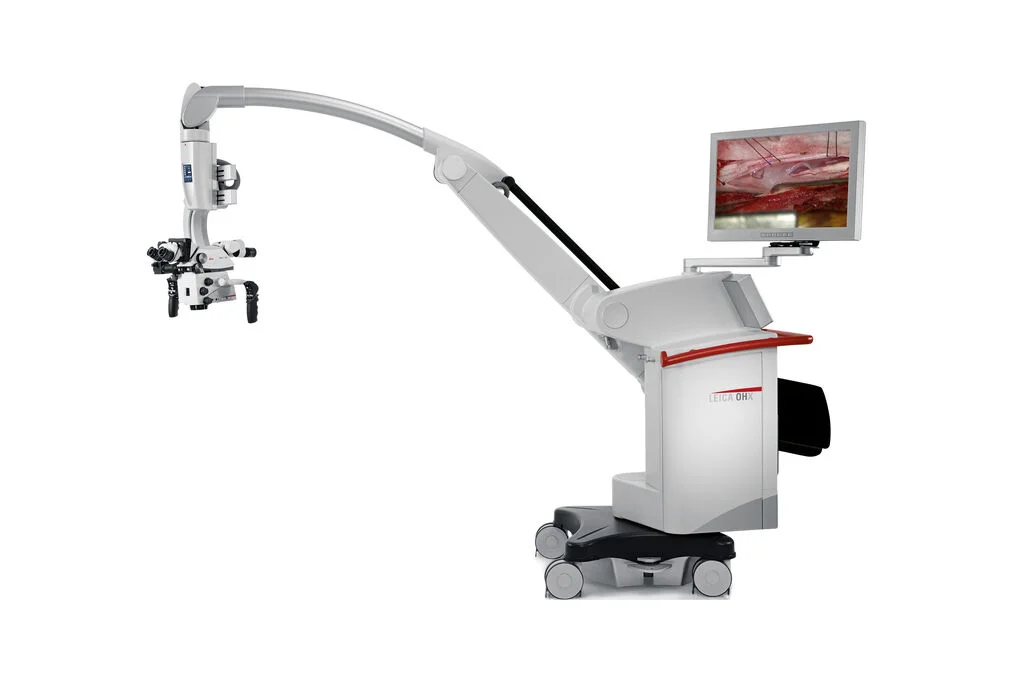
High-Definition Visualization

Magnified 3D view of neural structures

MIS Burrs

Targeted bone removal with specialized instruments

Muscle-Sparing Approach

Muscles are separated, not cut

The Procedure

1

Small Incision

A small incision is made in the lower back

2

Muscle Separation

Muscles are gently separated rather than cut to access the spine

3

Visualization

Endoscope or microscope provides magnified view of the surgical area

4

Decompression

Bone and soft tissue compressing the nerve are carefully removed

5

Confirmation & Closure

Surgeon confirms nerve has adequate space and closes the incision

Your Spine Specialist

Fellowship-trained neurosurgeon with expertise in minimally invasive spine surgery

Frequently Asked Questions

Endoscopic lumbar decompression is commonly used to treat lumbar spinal stenosis, disc herniation, radiculopathy (leg pain from nerve compression), and neurogenic claudication (pain with walking due to spinal narrowing). Your surgeon will review your imaging to confirm you are a suitable candidate.

Endoscopic lumbar decompression typically takes 1-2 hours depending on the complexity of the case and the number of levels being addressed. Because it is minimally invasive, many patients can go home the same day or after an overnight stay for observation.

Most patients can walk within hours of surgery. Light daily activities can often resume within 2-4 weeks, though this varies by individual. Physical therapy may be recommended to help strengthen the back and improve mobility. Full recovery typically takes 6-12 weeks depending on the extent of the procedure.

Because the procedure is minimally invasive, most patients experience less post-operative pain compared to traditional open surgery. Pain is typically well-managed with medications, and many patients report significant relief from their pre-operative symptoms within the first few weeks.

No referral is required. You can book a consultation directly with our spine specialist to have your condition assessed. Having a recent MRI is helpful but not mandatory—we can arrange imaging if needed.

Ready to Take the Next Step?

Schedule a consultation to discuss your treatment options.